







As the body matures, changes in bone density and muscle mass can occur naturally, yet these shifts sometimes accelerate into conditions like osteoporosis. Dual-energy X-ray absorptiometry, commonly known as a DEXA scan, has emerged as a primary tool in monitoring these physiological changes. By providing precise measurements of bone mineral density and body composition, these scans allow individuals and their healthcare providers to track health metrics over time. This data serves as a foundation for informed decision-making regarding one’s health.
What Is a DEXA Scan?
A DEXA scan is a specialized imaging test designed to measure bone mineral density. The technology uses two low-energy X-ray beams with different energy levels. This non-invasive procedure provides a T-score, which compares an individual’s bone density to that of a healthy 30-year-old adult. A score of -1.0 or above is generally normal, while scores between -1.0 and -2.5 indicate low bone mass, often referred to as osteopenia. A score of -2.5 or lower typically signifies osteoporosis. Beyond bone health, advanced DEXA technology assesses body composition. It distinguishes between soft tissue and bone tissue.
How Can a Scan Help?
Doctors frequently recommend DEXA scans for women aged 65 and older and men aged 70 and older. These age groups face higher risks of bone density loss due to hormonal changes and natural aging processes. Postmenopausal women experience a rapid decline in estrogen, a hormone that protects bones, making them primary candidates for screening.
Younger individuals may also need screening if specific risk factors exist. A history of fractures from minor impacts and long-term use of steroid medications can necessitate earlier testing. Understanding the ratio of muscle to fat may help in tailoring fitness regimens to prevent muscle loss while shedding fat. This specific application supports aging adults who wish to maintain strength and stability to prevent falls.
What Should You Expect?
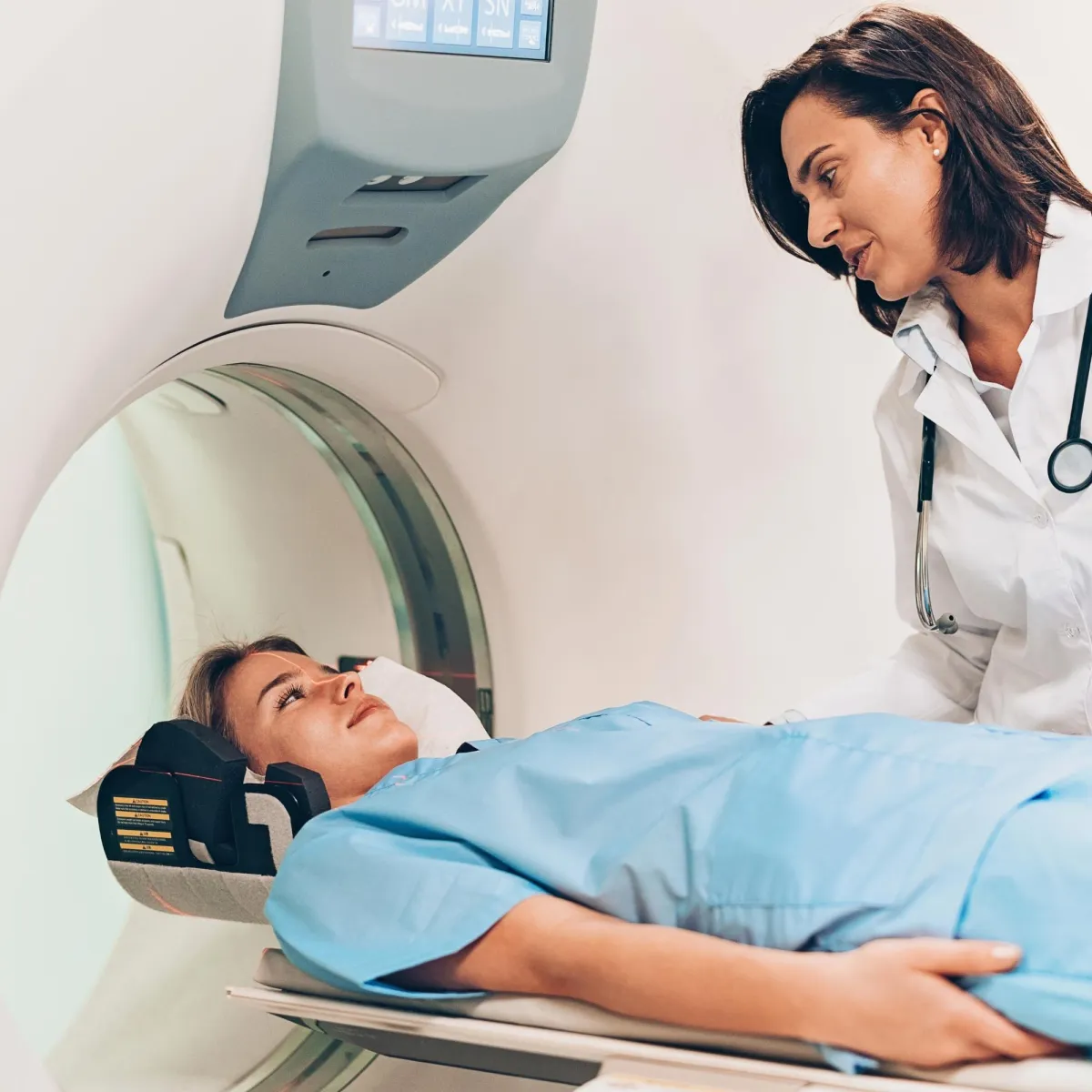
Undergoing a DEXA scan is a quick and painless process. No injections or anesthesia are required. Patients lie still on an open, padded table, then a scanning arm passes over their body. The device does not touch the patient, and the open design may help avoid feelings of claustrophobia.
Preparation is minimal. Patients can eat normally on the day of the exam, but should avoid taking calcium supplements for at least 24 hours prior. Loose, comfortable clothing without metal zippers, buttons, or belts is best. In some cases, patients may wear a hospital gown to prevent image artifacts caused by clothing fasteners.

Radiation exposure during the procedure is extremely low. Once the scan concludes, a radiologist analyzes the images and compiles a report. This report details bone density measurements and body composition data, which the referring physician then reviews with the patient.
Seek Professional Radiology Services
Interpreting the results of a DEXA scan requires specialized medical knowledge. While the scan provides raw data, a qualified radiologist or physician must analyze these numbers within the context of a patient’s full medical history. They look for trends over time to determine the best course of action. Regular monitoring through professional radiology services allows for the early detection of subtle changes in bone structure. This proactive approach enables the implementation of preventative strategies before fractures occur.